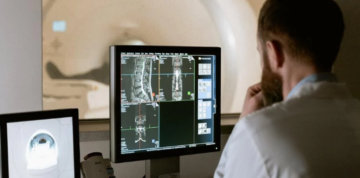
Medical imaging plays a pivotal role in modern healthcare, revolutionizing how medical professionals diagnose and treat many conditions. It allows clinicians to peer inside the human body without invasive procedures, aiding in early detection, precise diagnoses, and informed treatment planning. This article delves into the world of medical imaging equipment, tracing its evolution from the discovery of X-rays to cutting-edge 3D imaging technologies while also exploring the future of diagnostics.
The Birth of Medical Imaging
The origins of medical imaging can be traced back to the serendipitous discovery of X-rays by Wilhelm Conrad Roentgen in 1895. This revolutionary breakthrough revealed that X-rays could penetrate soft tissues but not dense structures like bones, creating a stark contrast in images. Almost immediately, X-ray imaging found applications in medicine.
X-ray Imaging
X-ray imaging is based on the principle of electromagnetic radiation. As X-rays traverse through the body, their absorption varies accordingly, creating shadows that can be captured on X-ray film or digital detectors. Dense structures, such as bones, appear white, while softer tissues appear in shades of gray.
Despite numerous technological advancements in medical imaging, X-rays continue to be a cornerstone in diagnostic radiology. They are instrumental in diagnosing bone fractures, detecting foreign objects, and evaluating pulmonary conditions, such as pneumonia or lung cancer. Portable X-ray machines have also become essential in emergency and critical care settings.
Advancements in Medical Imaging
Advancements in medical imaging have transformed the healthcare field by providing more precise, less invasive, and increasingly informative methods for diagnosing and monitoring medical conditions. These advancements have greatly improved patient care, treatment planning, and medical research. Here are some key advancements in medical imaging:
- Magnetic Resonance Imaging (MRI): MRI is a non-invasive imaging method that employs strong magnetic fields and radio waves to produce intricate images of the internal structures within the body. Unlike X-rays or CT scans, MRI avoids employing ionizing radiation, which enhances patient safety. MRI is especially valuable for visualizing soft tissues like the brain, spinal cord, muscles, and organs. Recent advancements in MRI technology have led to faster imaging, higher resolution, and specialized MRI techniques for specific applications, such as functional MRI (fMRI), for studying brain activity.
- Computed Tomography (CT) Scanning: CT scans merge X-ray technology with computer processing to generate cross-sectional images of the body. Advances in CT imaging have resulted in faster scan times, reduced radiation exposure, and improved image quality. Dual-energy CT can provide additional information about tissue composition, while spectral CT can differentiate materials based on their atomic arrangement. These advancements are crucial for diagnosing various conditions, including cancer, cardiovascular diseases, and trauma injuries.
- Ultrasound Imaging: Ultrasound, which uses high-frequency sound waves to create images, has significantly improved in recent years. Portable handheld ultrasound devices have become more accessible, enabling point-of-care imaging in various medical settings. Additionally, 3D and 4D ultrasound technology offers a more comprehensive view of developing fetuses, making it an essential tool in obstetrics. Innovations in ultrasound also include better image quality and improved image processing techniques.
- Positron Emission Tomography (PET) Scans: PET scans have evolved with the development of more precise radiotracers and hybrid imaging systems. Integrated PET-CT and PET-MRI scanners provide anatomical and functional information, improving the accuracy of cancer staging and disease localization. New radiotracers are continually being developed to target specific molecular processes, allowing for early disease detection and monitoring of treatment response.
- 3D and 4D Imaging: Three-dimensional (3D) and four-dimensional (4D) imaging technologies have become essential in various medical fields. 3D imaging provides volumetric data that aids in surgical planning, dental implant placement, and orthopedic procedures. Meanwhile, 4D imaging adds the dimension of time, allowing clinicians to monitor dynamic processes like fetal development and cardiac function in real-time. These advancements enhance precision and improve patient outcomes.
Artificial Intelligence (AI)
Artificial intelligence (AI) has emerged as a transformative force in medical imaging, bringing about significant advancements in image analysis and personalized medicine. These AI applications are revolutionizing how we approach diagnostics and treatment in healthcare.
AI in Image Analysis
One of AI’s most notable medical imaging contributions is its image analysis prowess. AI algorithms, particularly deep learning models, have demonstrated remarkable capabilities in interpreting medical images with a level of precision and efficiency previously unattainable.
- Improved Accuracy: AI systems can detect subtle abnormalities, such as early-stage tumors or microfractures, in medical images with high accuracy. This has the potential to lead to earlier diagnoses and more effective treatments.
- Speed and Efficiency: AI can process large volumes of medical images in a fraction of the time it would take a human radiologist. This accelerated analysis can significantly reduce the time it takes to deliver results to patients, which is particularly crucial in emergency cases.
- Automation: Routine tasks like image segmentation, organ recognition, and anomaly detection can be automated with AI, allowing radiologists and healthcare professionals to focus on more complex aspects of patient care.
- Quantitative Analysis: AI can provide quantitative data from medical images, enabling more precise monitoring of disease progression and treatment effectiveness. This is especially valuable in conditions like cancer, where tracking changes over time is critical.
- Pattern Recognition: AI algorithms excel at recognizing patterns and can identify trends and associations in medical data that may not be readily apparent to human observers. This can aid in research and the discovery of new diagnostic markers.
Challenges and Ethical Considerations
While medical imaging has significantly advanced healthcare, it also brings challenges and ethical considerations that must be carefully addressed.
Radiation Exposure
While medical imaging has countless benefits, it raises concerns about radiation exposure. Techniques such as X-rays and CT scans involve ionizing radiation, which can pose health risks with repeated use. Therefore, careful dose management is crucial.
Data Privacy and Security
The digital nature of medical images and the integration of AI bring forth challenges related to data privacy and security. Protecting patient data from breaches and ensuring the ethical use of AI algorithms are ongoing concerns in the field.
Future Directions in Medical Imaging
The future of medical imaging holds exciting possibilities. Researchers are exploring novel imaging modalities, such as hyperspectral imaging and photoacoustic imaging, which could provide unprecedented insights into tissue composition and function. Furthermore, the integration of AI and big data promises to revolutionize diagnostics, making it more precise and accessible.
Final Thought
In the ever-evolving world of medical imaging, A+ Medical Company, Inc. is your trusted partner for all your equipment and parts needs. Whether in a hospital, clinic, or independent healthcare practice, they will support you with top-quality new, used, and refurbished medical imaging equipment. As a leading provider in the field, they’re committed to helping you stay at the forefront of healthcare technology. Explore what they can offer, and be part of advancing healthcare worldwide. Visit aplusmedical.biz now to discover more.